

Volunteer/Request Ambassador
.svg)
FACTOR 2025


Biological Drivers of Osteosarcoma
View Speakers & Abstracts.png)
James Morrow, MD, PhD
Dana-Farber Cancer Institute

Dissecting the developmental origins of osteosarcoma
There has not been a significant change in the way that we treat osteosarcoma for more than 30 years. One of the primary reasons why it has been difficult to develop and test new therapies is that we as a community understand very little about the biology of osteosarcoma compared to other more common cancers. Remarkably, one of the things we don’t understand about osteosarcoma is what types of normal bone cells osteosarcoma comes f rom. We believe that this fundamental knowledge is key to understanding what drives osteosarcoma. In these studies, we have developed an experimental system that allows us to model bone development in the lab. For the first time this has allowed us to generate a reference map of normal bone cells to which osteosarcoma cells can be compared. Now that we have this system working, we can introduce DNA changes (mutations) that we see in osteosarcoma into normal bone cells. We find that when we introduce osteosarcoma mutations into developing bone cells we end up with new types of cells that don’t exist in normal development. We think these new cells are the first step in the development of osteosarcoma. Right now, we’re working to more fully understand how these abnormal cells come to be and how they evolve into tumors. In addition, when we compare our bone development reference maps to osteosarcoma tumors, we find that we learn a lot more about these tumors. For example, we have learned more about what causes osteosarcoma to spread, or metastasize. We hope that this knowledge will help us find weaknesses in osteosarcoma cells that we can exploit to create new therapies.
Kelly Gutpell, MBBCh, PhD
Nationwide Children's Hospital
Characterization of the anchor cell in preclinical models of metastatic osteosarcoma
Overall survival for patients with metastatic pulmonary osteosarcoma remains poor, and there is an urgent need for new therapeutic strategies to improve outcomes for these young patients. Metastasis to the lung is an inefficient process and previous studies have shown that a small population of tumor cells plays a crucial role in remodeling lung tissue to establish, or “anchor ”, the metastatic niche. These anchor cells are a potentially novel target, but they remain poorly understood at the gene expression level. This study aims to identify and characterize these anchor cells by analyzing gene expression at the single-cell level using RNA sequencing. Additionally, we will use lineage tracing barcodes to determine whether the anchor cell is clonally distinct f rom the rapidly dividing growth cells that give rise to metastatic lesions. This study will provide new insights into the biology of osteosarcoma metastasis and identify a potentially novel target during tumor evolution that could drastically impact the lives of young people with this disease.
Mathieu Epinette, BS
Koch Institute for Integrative Cancer Research at MIT
Heterogeneous and ongoing genomic rearrangements in a murine osteosarcoma model parallel human disease
Advancing a new drug or therapy to human trials requires a huge investment of time and money. In rare diseases like osteosarcoma, there are few chances to try new therapies, making it crucial to take our best shot when we can advance a new treatment. Preclinical models that faithfully reproduce human disease enable us to test therapies and prioritize for human study. Models range from cells derived f rom patient tumors grown in a dish to animals genetically engineered to get OS. Each has strengths and weaknesses that make them right for certain applications. However, in all cases, it is crucial to understand what aspects of OS a model recreates and where it differs. To study how the immune system interacts with OS, we use a mouse genetically engineered to develop OS. Unlike models that transplant human tumors into mice, these mice have a normal immune system. Original reports of this model describe how it reproduces the microscopic and clinical aspects of the disease, but it remains unknown whether it has the same genetic changes. Human OS is characterized by a process called chromothripsis, in which one or multiple chromosomes are broken apart into many tiny pieces and reassembled. This results in genes breaking apart and moving to new places in the genome. In the reassembly process, some genes are lost while others are accidentally copied too many times. Both types of changes likely contribute to the development of OS. To understand whether the mouse model of OS undergoes chromothripsis, we performed detailed gene sequencing of mouse leg tumors and, when the cancer had spread, f rom mouse lungs. From these data, we were able to see that many of the mouse tumors undergo complicated genomic rearrangements that look like chromothripsis. Furthermore, we found that this results in extra copies of genes known to cause human OS (Myc) and loss of genes that normally prevent OS (Pten). We have subsequently confirmed many of these changes on the protein level as well and are in the process of testing whether they contribute to the progression of OS. While many mouse models of cancer are genetically much simpler than their human equivalents, this mouse OS model faithfully reproduces the genetics seen in human OS. This makes it a great system for developing new ideas for therapies and ultimately for testing those therapies before they enter human clinical trials.
Sowmya Ramesh, PhD
Johns Hopkins University
Sensory neurons regulate osteosarcoma disease progression
Bone pain is a hallmark of bone cancers, including osteosarcoma (OS), mediated by peripheral neurons. However, the roles of tumor-associated sensory neurons in OS beyond pain perception remain poorly understood. To investigate their regulatory functions, a chemical-genetic approach was employed in mice carrying a knock-in allele for TrkA to perturb sensory nerve innervation during OS growth and progression. TrkA inhibition in these transgenic mice significantly reduced sarcoma-associated sensory innervation and vascularization, decreased tumor growth and metastasis, and extended overall survival. In a translational effort, we leveraged FDA-approved bupivacaine liposomes which led to significant reductions in tumor growth, blood vessels, as well as alleviation of pain. These data suggest that interventions to prevent pathological innervation of osteosarcoma represent a novel adjunctive therapy to improve clinical outcomes and survival.
Kevin Jones, MD
University of Utah, Huntsman Cancer Institute
Epigenetics and osteosarcoma oncogenesis
The product of osteosarcomagenesis, a full-fledged osteosarcoma, has been very clearly defined as long as physicians have been looking at tissue sections on glass slides under light microscopy: pleiomorphic (also spelled pleomorphic, meaning many shaped) cells are intimately associated with and apparently generating osseous (bony) extracellular matrix. Some have therefore used the more specific moniker or osteogenic sarcoma. Put simply “ugly cells making osteoid” defines an osteosarcoma. The ugliness of those cancer cells refers to the fact that the nucleus and cytoplasm of each cancer cell differs—often wildly—f rom adjacent cancer cells in the same microscopic tissue section f rom the same tumor. This can also be referred to as intratumoral and intercellular heterogeneity. That definition has remained consistent, even as molecular features of osteosarcoma have been resolved with increasingly sophisticated tools. Osteosarcoma was central to the initial recognition that cancer of most types has genetic origins. How we defined those genetic origins came initially f rom inherited conditions running in families, especially Li Fraumeni syndrome, congenital bilateral retinoblastoma syndrome, and Rothmund-Thomson syndrome. Eventually, the genes, whose mutations associated with each of these conditions were identified in the human genome, even to the point that the specific sequence aberrations that characterized the common mutations became resolved at the base-pair level. Eventually, two of these genes were recognized as disrupted in both the osteosarcomas associated with their respective syndromes, as well as in sporadic osteosarcomas that develop in individuals with no family history of osteosarcoma or even other cancers. However, also recognized as profiling became more deeply penetrant into osteosarcoma genomes was the observed molecular mayhem, the chromothripsis, or shattering of chromosomes that consistently characterizes them. As mouse genetic modeling of osteosarcoma has also improved f rom transgenes through targeted disruption of specific tumor suppressor genes in cells along the osteoblast lineage, the field has learned that the critical threshold that must be passed is the genome scrambling itself, whether permitted by the silencing of specific genes, or accelerated by the application of ionizing radiation, DNA damaging chemicals, or genetic drivers of DNA damage accumulation. The new goal of the field is to identify what epigenetic circumstances enable or generate the genetic smashing of the genome that still generates osteosarcoma cells that appear to have bizarrely many-shaped nuclei with highly variable genetic content even among the cells within a single cancer.
Collaborative Data Ecosystems
View Speakers & Abstracts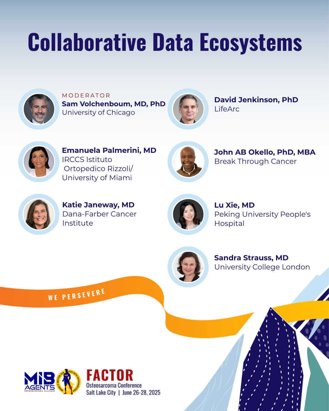
Katie Janeway, MD
Dana-Farber Cancer Institute
Directly engaging participants in rare cancer research is feasible: The Osteosarcoma Project
Background and purpose: Osteosarcoma (OS) is a sarcoma with a complex genome for which there has been limited progress in identifying new treatments and improving outcomes. While slow progress is partially due to insufficient genomic characterization, generating large genomic datasets has been challenging because this is a rare cancer. The OS Project uses various approaches to directly engage pediatric and adult participants with OS in genomics research. Methods: Working with patients and advocates at the design stage, we created a website (OSProject.org) where patients register and consent to participation. Any patient with OS living in the United States or Canada is eligible. Participant outreach approaches include partnership with advocacy organizations, webinars, social media posts, meeting presentations, stakeholder and physician engagement committees, and direct mailings. Blood and saliva are collected directly f rom consented participants by mail, archival FFPE tumor samples are obtained f rom pathology departments and medical records are requested f rom treating institutions. WES, WGS, DNA panel sequencing and RNASeq of tumor and germline (T/N) is performed. Results are shared with patient, advocacy, physician, and research communities in several ways. Individual participants receive a shared learning report describing the somatic variants identified in their tumor f rom T/N clinical WES and are offered clinical germline genetic testing and genetic counseling. Results: The study outreach team has participated in or led a total of 20 online and in person events not including social media posts or stakeholder meetings. So far, in 26 months 145 OS patients (ages 7-74y; median 20) have consented, and 76 tumor samples and 127 germline samples have been obtained f rom the consented participants. Analysis of the first 7 OS pts with T/N WGS demonstrated inactivation of TP53 (4 pts) and RB1 (2 pts), as expected f rom past OS cohorts. Conclusions: Using community partnerships and direct outreach to connect and engage with participants and a virtual consenting process for genomics research in a rare cancer is feasible. It is possible to obtain germline samples directly f rom about half of participants and archival tumor samples f rom treating pathology departments for about one third of participants consented in a direct-to-patient online genomics study. We have been able to utilize archival tumor samples to identify, with T/N WES/WGS, expected genomic events in complex genome cancers. Recruitment and sequencing are ongoing.
Emanuela Palmerini, MD, PhD
Sylvester Comprehensive Cancer Center, Miller School of Medicine, University of Miami
FOSTER (Fight OsteoSarcoma Through European Research) Consortium
The FOSTER Consortium (Fight OsteoSarcoma Through European Research) (TBD) is a Pan-European initiative dedicated to advancing research and improving outcomes for patients with osteosarcoma, the most common bone cancer in adolescents and young adults. With over 350 members across 20 countries, the consortium fosters collaboration among researchers, clinicians, and patient/parent advocates to address critical gaps in translational research, clinical trial access, and long-term survivorship. Organized into nine work packages, FOSTER supports biobanking, preclinical modeling, and the development of innovative therapeutic strategies to all patients with osteosarcoma. In a significant milestone, the consortium was awarded more than €7,600,000 through the ATTRACT* Call to investigate maintenance therapy with cabozantinib (FOSTER-CabOs trial), in partnership with Ipsen, in a randomized clinical trial enrolling all newly diagnosed patient with osteosarcoma in Europe. The FOSTER – CabOS trial is the first randomised question of the FOSTER-Evolving Study Platform registering all patients at diagnosis for biology studies, with initial financial support f rom Rising Tide. The FOSTER – CabOS trial will support a comprehensive clinical and translational research program aimed at evaluating the efficacy and safety of cabozantinib as a maintenance strategy in osteosarcoma, potentially offering a new therapeutic avenue for patients with limited treatment options.
Lu Xie, MD
Peking University People's Hospital
Collaborative data ecosystems in mainland China
To introduce the Collaborative Data Ecosystems panel in the mainland of China epecial for osteosarcoma. There are many sarcoma centers in the mainland of China, most of which are now still supervised by orthepedic oncologsts. This presentation is planned to introduce our current status as well as the appropriate way for us to communicate and coroperated in the future under current local policy.
David Jenkinson, PhD
LifeArc
Pediatric Bespoke Therapeutic Discovery Workshop on Osteosarcoma - Community Priorities
The Pediatric Bespoke Therapeutic Development Workshops (PBTDWs) are a series of workshops focussing on specific childhood cancers, run by LifeArc in collaboration with the Innovative Therapies for Children and Adolescents with Cancer (ITCC), Cancer Research UK and disease specific foundations. By convening world-leading experts, they aim to capture our current understanding of specific childhood cancers and explore potential targets that may be useful for new therapeutic discovery. They also look at the main clinical questions in the disease. This talk will cover the outcomes of the osteosarcoma workshop; the prioritisation of the targets discussed and the clinical opportunities around combinations of DNA damage response inhibitors, available in adult cancers that might be repurposed in osteosarcoma. It will also touch on the potential of targeting a protein called MYC, which is f requently overexpressed in poor prognosis disease.
John B.A. Okello, PhD, MBA
Break Through Cancer
Team DEFIANT: A Radical Collaboration Framework to Accelerate Therapeutic Discovery in Osteosarcoma
Osteosarcoma remains one of the most difficult pediatric malignancies to treat, due in part to its complex genomic landscape, lack of biomarker-guided therapies, and limited access to coordinated clinical and biological data. At the MIB FACTOR 2025 conference, BTC introduced their upcoming launch of Team DEFIANT (DEFIning ACTioNable Targets for Osteosarcoma)—a new $15 million TeamLab supported by Break Through Cancer (BTC) and other partners to be announced. This initiative will formally launch in Fall 2025 and represents one of the largest osteosarcoma-focused research investments to date. Team DEFIANT integrates BTC’s radical collaboration model, which unites multi- institutional investigators and supports real-time data sharing, cross-species discovery, and rapid translational learning. The effort is structured around two interdependent pillars: 1) Integrated Trial Framework: A unified human-canine trial ecosystem that incorporates advanced molecular diagnostics, surrogate endpoints, and minimal residual disease tracking, to accelerate therapeutic evaluation and clinical readiness. 2) Target Discovery Engine: A cross-disciplinary effort to aggregate and analyze existing and newly generated osteosarcoma datasets—including genomic, transcriptomic, spatial, and single-cell data—from over 600 human and canine cases. This pillar will focus on identifying and prioritizing actionable targets such as cell surface antigens and chromosomal instability signatures. Led by scientific collaborators from BTC’s founding institutions—including MSK, MD Anderson, DFCI, Johns Hopkins, MIT—as well as Stanford, UCSF, Tufts, and UBC, Team DEFIANT represents a novel, high-trust model for rare cancer research. The initiative is designed to serve as a national proof-of-concept for rapid, biomarker- driven innovation in osteosarcoma and is a key step toward BTC’s 2034 vision: to make real-time, biomarker-informed care standard for all patients with this disease.
Sandra Strauss, MD, PhD
UCL Cancer Institute
ICONIC: Improving outcomes through collaboration in osteosarcoma
Osteosarcoma (OS) is the most common bone cancer that affects children and young people, as well as some older adults. Treatment is challenging often involving traumatic surgery and intensive chemotherapy that is toxic with significant short- and long-term side effects. With no significant advances in treatment for the last 20 years and poor outcomes particularly for those with metastatic disease at diagnosis and relapse disease, there is an urgent need to better understand the biology of osteosarcoma and identify new treatments for OS patients and gain an insight into delays in diagnosis and poor patients experience. The ICONIC study, generously funded by the Bone Cancer Research Trust, was established to bring about a step change in how we conduct collaborative OS research in the UK and is a national prospective observational study that aimed to recruit all newly diagnosed OS patients of all ages to form a cohort with high-quality clinical data as well as imaging, blood and tissue samples to address clinical and biological questions prioritised by the community. Since opening, ICONIC has recruited 342 patients across 28 centres. An update on preliminary findings from the study will be presented.
Tumor Heterogeneity and Microenvironment Interactions
View Speakers & Abstracts
Greg Sawyer, PhD
Moffitt Cancer Center
3D patient avatars, cell therapy, and microscopic mayhem
Over the past century, imaging, drug discovery, and cancer biology have been locked in essentially two-dimensional systems due to the ubiquity and standardization of an extensive inf rastructure built around culture plates, slides, and imaging dishes. The inventions of scanning-confocal microscopy and multiphoton microscopy are poised to bring rapid, three-dimensional high- resolution characterization of tissues to medical research once the seemingly intractable problem of maintaining, handling, and working with functional, three- dimensional human microtissue specimens is solved. This presentation outlines a radical new approach to convey human biology to medical science and develop multi-organ systems as well as patient-specific models of disease in an accessible platform optimized for in situ studies of tissue function and dynamics with high- resolution microscopy. We will present in situ measurements of immunotherapy activity in 3D systems of patient derived micro-tumors with expanded cultures of TILs, PBMCs, and CAR T cells. These experiments will follow immune cell migration, killing, and tumor elimination using in situ scanning confocal microscopy, live-cell imaging techniques, astrophysics inspired AI tracking, spatio- temporal 3D cytokine measurements, and evolutionary dynamics modeling of immunotherapy.
Anjali Garg, PhD
Center for Cancer Research, National Cancer Institute, National Institutes of Health
Toward a greater understanding of shared molecular alterations in canine and human osteosarcoma: an update from the NCI DOG2 project
Objective: Clinical outcomes for osteosarcoma (OS) continue to stagnate despite decades of dedicated research toward understanding the inherent complexity of the disease. Like many cancers, OS lacks a clear set of genomic driving events and exhibits significant intra- and inter-patient heterogeneity, reflected in histologic patterns, variable immune infiltration and tumor microenvironment (TME) composition, and chaotic chromosomal events. Further, tumor-derived biomarkers such as % necrosis after neoadjuvant chemotherapy or MYC amplification may fail to accurately predict which patients are at highest risk for disease progression. Canine OS represents a common and valuable animal patient model of the human disease within which predictive biomarkers and novel therapeutic approaches can be discovered and tested within comparative oncology clinical trials. However, to have the highest translational impact for humans, tractable methods for canine patient stratification based on genomic features are needed. Methods: Leveraging biologic samples f rom canine trials, we used low-pass whole- genome sequencing in a cohort of canine OS patients (n = 261) to study copy number variations (CNV). From this same cohort we have matched mRNAseq data f rom a subset of n = 186 tumors and whole-genome bisulfite sequencing f rom n = 47. Results: CNVs are common in canine OS. Patterns of gains and losses in observed tumors are reflected in a panel of n = 27 canine OS cell lines that were developed by veterinary researchers across the US. CNVs involve most of the same genes that are altered in human OS, such as MYC, PDGFRa, PTEN, CDKN2A/B and ATRX. Specific CNVs carry prognostic value in a discovery cohort of 52 canine tumors, but additional work in a separate validation cohort of n = 209 tumors is underway to confirm these early findings. Our group has already established the prognostic value of TME subtype, with immune-enriched tumors having a more favorable outcome than without immune cell infiltration. This new analysis shows that primary tumor methylation status predicts outcome in canine OS, with hypomethylated tumors having a significantly decreased survival, which correlated with an immune-depleted TME. Conclusions: This integrated approach supports correlation between gene dose, methylation status, and TME subtype as well as a deeper understanding of the biology of the disease in dogs.
Yogesh Budhathoki, MS, PhD Candidate
Nationwide Children's Hospital
Project OsteoCAR: Building a comprehensive resource for cell type annotation and transcriptomic exploration of osteosarcoma
Our work aims to create OsteoCAR (Osteosarcoma Cell Type Annotation Reference), a reliable single-cell reference dataset incorporating a comprehensive understanding of heterogeneity within the primary and metastatic microenvironments across patients and animal models. The strength of this project lies in compiling, annotating, and analyzing an enormous cross-species dataset that we and others can leverage to explore specific aspects of osteosarcoma biology. Through a collaborative effort, we have assembled a unified dataset containing about 1,000,000 cells f rom human patients, patient-derived xenografts, key mouse models, and canine patients. The samples come f rom a diverse cohort of patients and animal models with primary and metastatic lesions. Utilizing various computational tools, our analysis pipeline first integrates the dataset per modal and tumor site. It then identifies and splits the tumor and stroma cells for downstream analysis, where we annotate the cell types at the granular level utilizing cell type markers, differentially expressed gene analysis, gene set enrichment analysis, and other relevant computational analysis. With these annotations, we have begun systematically evaluating the inter- and intra-tumor heterogeneity and the patterns of host cell composition and phenotype that characterize the primary and metastatic microenvironment across species. Our preliminary results have identified consistent patterns of intra- tumoral heterogeneity that are recurrent and predictable across biological contexts. Some tumor cell subpopulations have immature proliferative stem cell- like features, while others have more differentiated inflammatory or matrix- forming osteoblast- and fibroblast-like phenotypes. A population of tumor- associated macrophages with an osteoclast-like phenotype is shared among all tumors. Macrophages with scar-like phenotypes are much more prominent in lung lesions. We expect that this resource will be valuable for 1) defining osteosarcoma tumor cell subsets and creating a shared language for discussion of osteosarcoma intra- tumoral heterogeneity, 2) automated assignment of cell types in single-cell and spatial workflows, 3) studying tumor-host interactions within both primary tumors and metastatic lesions, 4) evaluating tumor cell plasticity and the evolution of malignant cells over time, both during metastasis and in response to therapeutic stresses, and 5) translation of single-cell biology across models and species.
Rebecca Makii, MS, DVM, DACVP
Colorado State University
A comparative understanding of oncogenic MYC signaling in the osteosarcoma metastatic tumor immune microenvironment
Osteosarcoma (OS) is a debilitating bone cancer that impacts both humans and dogs with similarly poor clinical outcomes. Canine OS occurs more f requently and has been used as a model to study human OS and accelerate treatment discoveries. MYC is a protein that influences a variety of cellular functions. Overexpression of MYC has been associated with poorer outcomes in human OS and has recently been described in canine OS. However, the mechanism by which MYC is associated with poor prognosis in OS is not understood. Recent data examining MYC overexpression in other cancers has demonstrated that MYC can alter the types of immune cells present within a tumor. This is primarily through increasing macrophages and decreasing T cells, which leads to local immunosuppression and worse outcomes. These findings can ultimately decrease response to immunotherapies. We therefore hypothesize that MYC is contributing to the overall immunosuppression found in metastatic OS through similar immune altering mechanisms. Using the dog as a naturally occurring model to study metastatic disease, we identified archived samples of OS lung metastases to evaluate MYC at the DNA, RNA, and protein level. We demonstrate that despite relatively normal or low levels of MYC DNA or RNA, there is increased MYC protein in a subset of our canine metastatic OS samples. We further show that samples with elevated MYC protein expression are associated with signals indicating a locally immunosuppressive metastatic environment. In human and canine OS cell culture models, we demonstrate effective targeting of MYC using a novel drug (Myci975). We show that this drug alters tumor cell survival and inflammatory chemical signaling. Therefore, targeting MYC may serve as a good strategy to increase efficacy of the currently available immunotherapies, which are often considered ineffective in OS. Additionally, metastatic OS MYC protein expression may have utility as a prognostic indicator for response to immunotherapy treatment.
Alexander Davies, DVM, PhD
Oregon Health & Science University
Dynamic tissue models reveal targetable mechanisms of single cell drug resistance in complex tumor ecosystems
Rawan Makkawi 1, Vaibhav Murthy 1, Jeremy Copperman 1, Alexander E. Davies 1,2,3
1 Cancer Early Detection Advanced Research Center, Knight Cancer Institute, Oregon Health and Science University, Portland, OR, USA.
2 Division of Oncological Sciences, Knight Cancer Institute, Oregon Health and Science University, Portland, OR, USA.
3 Department of Pediatrics, Oregon Health and Science University, Portland, OR, USA.
Individual cancer cell behavior results from the complex interplay of mutated or otherwise phenotypically reprogrammed cells with the host microenvironment. Our understanding of this process has often been inferred by collecting data at snapshots in time, limiting our ability to resolve tissue and molecular dynamics at the root of disease pathogenesis and drug response at the single cell-level. Our objective was to address this limitation by combining ex vivo approaches, fluorescent biosensors, and imaging techniques, into a unified live-cell dynamic model of cancer metastasis in the lung, capable of extracting quantitative spatiotemporal relationships and identifying causal links between tumor- host and tumor-tumor interactions underlying single cell fate. Using this model, the lungSITE (Serial Imaging of Tumor and microEnvironment), we found that metastatic osteosarcoma signaling dynamics were highly sensitive to positional and temporal variation in the physical microenvironment of the lung. Tumor cells at the host interface frequently displayed elevated signaling whereas cells in the tumor core displayed relatively low signaling activity. Initial single cell drug responses strongly correlated with position, whereby signaling 'high' host tissue interface cells persisted longer than cells in the core. Strikingly, a rapid adaptive response was observed in a fraction of interface cells following death of cells in the core, eliciting a drug resistance phenotype. We discovered that cell death in the core released tumor-derived growth factors into microenvironment. When combined with factors derived from the host, a resistance response was initiated that could be overcome by addition of receptor tyrosine kinase inhibitors. Together, these results demonstrate the utility of the lungSITE model to quantitatively measure tumor signaling dynamics and behaviors within the context of the lung metastatic niche. Data obtained from this model provided new insights into how spatial position and temporal response influence signaling dynamics, create heterogeneity, and can be targeted to overcome single cell drug response variation.
Mechanisms of Resistance and Evolution
View Speakers & Abstracts
Katia Scotlandi, PhD
IRCCS Istituto Ortopedico Rizzoli
Molecularly-driven information on patient-derived xenografts (PDX) and cell lines leads to ixabepilone repurposing for resistant osteosarcoma
Patients with highly aggressive, chemoresistant osteosarcoma (OS) continues to represent a population with unmet need. There has historically been a limited number of relevant models that well represent the heterogeneity of OS to be used for developing discovering efficient novel drugs. We have enlarged the already available collections of patient–derived xenograft (PDX) with 30 additional models that depict the epidemiologic, genetic, and biological features of OS and we have established six, new PDX-derived cell lines as a resource to generate solid preclinical findings to guide treatments for patients with these aggressive malignancies. Drug screening of 2800 FDA-approved compounds led to identification of five repurposed drugs, including ixabepilone that was selected as the most effective agent against highly aggressive OS with features of innate and/or acquired chemoresistance. The development of new drugs for rare tumors, such as OS, is a major challenge. Indeed, considering that the process of developing a novel drug to treat any kind of disease is typically laborious, costly, and failure-prone, development of new agents for rare tumors is particularly unappealing for large manufacturers. In addition, for the few agents that are developed, market prices are usually extremely high, resulting in reduced access for patients. In this context, drug reuse is an appealing and promising approach to developing medicines for rare tumors. By utilizing already available medications with deep knowledge of pharmacological characteristics, action mechanisms, and safety profiles, drug repurpose may indeed speed up the inclusion of novel drugs for the treatment OS patients. Our study supports the incorporation of ixabepilone, a drug with a manageable toxicological profile and approved for the treatment of resistant breast tumors, into treatment plans for chemoresistant OS.
Michael Lizardo, PhD
BC Cancer
Inhibition of the mitochondrial oxidative stress response in metastatic osteosarcoma cells prevents growth in murine lung tissue
Treatment options for osteosarcoma (OS) patients with metastatic disease are currently lacking. There is a dire need to develop newer therapies that specifically prevent or inhibit the growth of lung metastases. We therefore set out to study how metastatic OS cells adapt and grow in experimental conditions in a petri dish that mimic the harsh conditions of the lung environment. More specifically, we wanted to determine how the mitochondria (the powerhouse of the cell) of metastatic cells change and adapt to the harsh environment of the lung. From previous research, an antioxidant protein called nuclear factor erythroid 2–related factor-2 (NRF2) is upregulated in metastatic OS cells in the lung, and we know NRF2 can increase the abundance of peroxiredoxin-3 (PRDX3), an antioxidant enzyme found in mitochondria. We therefore tested whether an antibiotic compound called thiostrepton (TS), which is known to inhibit the activity of PRDX3, can inhibit the growth of metastatic OS cells. Under experimental conditions that mimic the lung environment, we discovered that TS disrupted the mitochondria of metastatic OS cells. Moreover, TS-treatment inhibited the growth of metastatic OS cells inside mouse lung tissue maintained in a culture dish. These findings suggest that targeting the adaptive response of mitochondria in metastatic OS cells with a drug like TS may have high therapeutic potential for OS patients.
Shahana Mahajan, PhD
Hunter College, City University of New York
Repurposing drugs for osteosarcoma
The lack of targetable mutations is osteosarcoma contribute to poor treatment outcomes. Drugs that alter metabolic profile or molecular pathways that support cell growth may offer an alternative approach to control osteosarcoma growth. Research in our lab has shown Riluzole, an FAD approved drug, as an effective agent that inhibited growth, induced cell death, and prevented metastatic behavior in two metastatic osteosarcoma cell lines. Given the substantial genetic heterogeneity in osteosarcoma, studies on the efficacy of Riluzole in diverse osteosarcoma will be an asset in developing preclinical studies. Toward this goal, we investigated the effects of Riluzole on 11 osteosarcoma cell lines derived f rom primary or metastatic tumors of mouse or human origin and on five independent patient-derived xenograft (PDX) tumor cell lines. We found that Riluzole suppresses cell growth and inhibits cell invasion in most of the osteosarcoma cell lines, including PDX cell lines. The bioavailability of Riluzole is affected by oxidative metabolism by cytochrome P450 enzyme, CYP1A2, and the expression level of the enzyme varies significantly in the human population leading to effective dosage variability. Therefore, we tested the efficacy of prodrugs of FC3423 and Troriluzole in osteosarcoma. Our data demonstrates that FC3423 and Troriluzole, are significantly more effective in osteosarcoma growth inhibition both in vitro and in vivo.
Sasha Blay, PhD Candidate
The Hospital for Sick Children
Harnessing mutational signatures to pinpoint the development of chemoresistance in metastatic and relapsed osteosarcoma
Survivors of pediatric cancer face lifelong battles with severe morbidities, which includes a significant risk of recurrence. Mutational signatures are patterns of somatic mutations in the cancer genome with specific etiologies. Recent work has identified mutational signatures linked to chemotherapy agents in the genomes of relapsed pediatric patients, and our ability to detect this “battle scar ” signifies chemoresistance. However, there is still much to learn about how chemoresistance develops pediatric patients, including when and where the effects of chemotherapy are felt in the cancer genome. I thus developed a computational pipeline to combine mutational signature analysis with clonal evolution reconstruction to elucidate changes in mutational processes throughout disease progression. I then used this pipeline to analyse 1,743 pediatric tumour genomes f rom 10 pediatric cancer datasets, including 101 osteosarcoma tumour samples. I detected mutational signatures linked to 4 chemotherapy drugs: temozolomide, platinum-based agents, fluorouracil, and thiopurine. Of 235 samples with confirmed exposure, 37.9% displayed one or more therapy-associated signatures. When mutational signatures were examined in the context of the phylogenetic tree, I identified specific subpopulations with these chemotherapy signatures, demarking subpopulation-level resistance. In cases with multiple sequenced tumour samples, resistant subpopulations in recurrences and metastases were traced back to ancestors present in primary tumors at the time of diagnosis, suggesting certain subpopulations possessed the ability to withstand chemotherapy-induced pressures f rom an early stage and then expanded following treatment. Finally, osteosarcoma with pulmonary metastases showed platinum signatures across all sites, yet the signatures arose independently in separate subclones. The pervasive nature of this signature indicates a convergence to a resistant phenotype, and in osteosarcoma, this occurs later and independent to metastatic mechanisms. This research offers valuable insights for identifying pediatric patients at greater risk of recurrence and aiding the development of more effective treatment plans to enhance quality of life for cancer survivors.
Chelsey Burke, MD
Stanford University School of Medicine
Understanding tumor evolution and mechanisms of resistance in osteosarcoma
Outcomes for children with cancer have steadily improved over the past several decades; however, survival rates for children with advanced osteosarcoma have not improved significantly in the last 30 years. Less than 20% of osteosarcoma patients with metastasis survive long-term, largely due to the lack of understanding of tumor evolution and the mechanisms of treatment resistance. Our study aims to address these gaps by using patient-derived xenograft models to study osteosarcoma's evolution, focusing on the mechanisms driving resistance and metastatic progression. Through a novel systems biology approach, we will identify key proteins, known as "Master Regulators" (MRs), that drive osteosarcoma growth and metastasis. We will utilize single-cell sequencing techniques to study the tumor at a deeper level, revealing previously hidden insights into the genetic and molecular landscape of osteosarcoma. By identifying drugs that target these MRs, we aim to overcome the drug resistance that develops in progressive osteosarcoma. By prioritizing clinically applicable drugs, we anticipate generating translational results that can be used to inform future pediatric clinical trials. This innovative approach to studying the evolutionary dynamics of osteosarcoma, paired with advanced computational methods, promises to uncover critical mechanisms behind drug resistance and malignant progression. The knowledge gained f rom this research will enable earlier therapeutic interventions, ultimately preventing metastasis and improving survival outcomes for children and adolescents with high-risk osteosarcoma. Our goal is to bring these discoveries into clinical practice, addressing an urgent unmet need and transforming the role of precision medicine in pediatric oncology.
Precision Medicine
View Speakers & Abstracts.png)
Amanda Marinoff, MD
UCSF
Clinical biomarkers for osteosarcoma stratification(cBOSS): Insights froman international working group
Osteosarcoma is a biologically heterogeneous disease, yet current clinical management remains one-size-fits-all. Advances in molecular profiling have identified potential biomarkers that could refine risk stratification and guide therapeutic decision-making, but none have been integrated into standard care. The Clinical Biomarkers for Osteosarcoma Stratification (cBOSS) working group convened an international panel of experts to evaluate and prioritize the most promising molecular biomarkers for clinical implementation. We systematically reviewed the prognostic significance and level of evidence supporting key candidates, circulating tumor DNA (ctDNA) dynamics, copy number alterations, tumor microenvironment composition, and multi-omic classification strategies. Consideration was given to their reproducibility, biological relevance, and feasibility for integration into clinical trials. Our findings highlight ctDNA burden has demonstrated independent prognostic value across multiple studies, warranting standardization of methodologies for prospective validation. Additional promising approaches, copy number alterations, transcriptomic classifiers, genome-wide loss of heterozygosity, proteomic and epigenetic f rameworks, and tumor immune profiling, require further correlation with clinical outcomes and assessment of biological overlap. This presentation will discuss the current landscape of osteosarcoma biomarker development, the challenges of translating molecular discoveries into clinical practice, and the next steps for prospective validation in future trials. By integrating molecular and clinical risk factors, we aim to move toward a precision medicine f ramework that enables risk-adapted, biology-informed treatment strategies for osteosarcoma.
David Shulman, MD
Dana-Farber Cancer Institute
Prospective evaluation of ctDNA among patients with localized osteosarcoma identifies patients at high-risk of disease relapse: A report from the LEOPARD study
Background: Outcomes for patients with osteosarcoma have not improved in decades and a lack of tools to understand risk of relapse has impaired our ability to develop new strategies to treatment. Prior retrospective analyses demonstrated that elevated pre-treatment ctDNA was associated with an increased risk of relapse. The primary objective of the LEOPARD study (Liquid biopsy in Ewing sarcoma and osteosarcoma as a prognostic and response diagnostic) localized osteosarcoma cohort was to prospectively study ctDNA in patients with localized osteosarcoma. Methods: The LEOPARD study is a prospective, multicenter ctDNA study for patients with bone sarcomas. Patients enrolled through one of 12 primary study sites, or samples were obtained f rom patients enrolled to the Children’s Oncology Group Project EveryChild (ABTR18B1-Q). Patients were 1-50 years of age at enrollment, had a histologic diagnosis of unresected high-grade osteosarcoma and were planned to receive standard chemotherapy as per COG AOST0331 (primary study centers only). We utilized a validated ctDNA assay and planned to enroll a minimum of 113 patients. Results: Between October 1, 2017 and March 3, 2023, 138 patients with osteosarcoma met eligibility and enrolled. 133 patients had an evaluable pre- treatment ctDNA measurement. At diagnosis, 84 (63%) patients had detectable ctDNA and the median ctDNA level was 5.3% (range: 0-43.1%). Baseline detectable ctDNA burden (≥3%) was associated with an increased risk of relapse compared to undetectable ctDNA (2-year EFS: 56% [95% CI 45-68%] vs. 88% [95% CI 78-98%], respectively; P<0.0001). A similar pattern was seen using a cut point of ≥5% (53% [95% CI 41-67%] vs. 83% [95% CI 74-94%], P<0.0001), or ≥15% (37% [95% CI 23-60%] vs. 76% [95% CI 68-86%], P=0.0004). Analysis of on-treatment ctDNA is ongoing as well as associated molecular biomarkers. Conclusions: These findings prospectively validate prior results associating ctDNA detection with inferior survival in patients with localized osteosarcoma.
Masanori Hayashi, MD
University of Colorado
Correlative studies from NPCF Phase II trial of gemcitabine and nabpaclitaxel for recurrent osteosarcoma
The combination of gemcitabine and docetaxel is often used to treat patients with recurrent osteosarcoma. Nab-paclitaxel has preclinical activity against osteosarcoma and is potentially less myelosuppressive than docetaxel. The National Pediatric Cancer Foundation conducted a prospective multi-institutional phase II trial combining gemcitabine and nab-paclitaxel for patients with recurrent osteosarcoma. Liquid biopsy samples were collected every two cycles, and circulating tumor cell and circulating tumor DNA burden were assessed. Eighteen patients were enrolled, and all patients had detectable CTCs, which detected progressive disease in most patients. Circulating tumor DNA was detected in ten patients, and common genetic amplifications in osteosarcoma, such as MYC, Rad21, CCND3 were detected.
Emanuela Palmerini, MD, PhD
Sylvester Comprehensive Cancer Center, Miller School of Medicine, University of Miami
Prognostic and mifamurtide-related signatures for localized osteosarcoma: an ISG/OS2 trial correlative study of tumor immune microenvironment (TME)
Background: Two clinical trials in Italy (ISG/OS-2) and Spain (GEIS-33) focused on patients with non-metastatic osteosarcoma. These trials used a risk-adapted approach based on the presence of a protein called Pgp. Patients with Pgp-positive tumors received chemotherapy plus mifamurtide, an immune-activating drug, while Pgp-negative patients received chemotherapy alone (Palmerini E et al, JCO 2025 in press). The main goal of this study was to find gene-based markers in the tumor immune microenvironment (TME) that could predict patient outcomes and response to mifamurtide. Methods: Researchers analyzed RNA from tumor samples of 62 patients using a NanoString immune profiling panel. Results: A 21-gene signature was identified that could classify patients into high-risk (Hi-R) and low-risk (Lo-R) groups. The 5-year overall survival was 47% for Hi-R and 92% for Lo-R patients (p = 3e-06). Lo-R tumors had more CD8 T-cells, T-regs, and activated NK cells, and fewer CD4 T-cells—suggesting a more favorable immune environment. This 21-gene signature was validated in two independent datasets (Target-OS TCGA and GSE33382). Among patients treated with mifamurtide, a separate 31-gene signature was found to predict better survival and event-free survival (p = 1e-09 and p = 3e-06, respectively). Both gene signatures were independently linked to survival in multivariate analysis. Conclusions: The 21-gene signature can help predict prognosis in osteosarcoma, regardless of treatment. The 31-gene signature may help identify which patients will benefit from mifamurtide. Only 5 genes overlapped between the two signatures, suggesting mifamurtide works through a unique immune pathway. These findings support the need for a multicenter international trial to validate the 31-gene signature. This could lead to more personalized treatment, avoiding unnecessary 9-month mifamurtide regimens in patients unlikely to benefit. This research highlights the potential of using immune gene profiling to guide treatment decisions in osteosarcoma.
Matthew Dietz, DO, MSEd
University of Utah/Primary Children's Hospital
Prognostic significance and microenvironment influence of cMYC in osteosarcoma
The identification of biomarkers that predict a patients chance of survival is necessary to develop risk stratified treatments for patients with osteosarcoma. The gene cMYC is abnormally over-expressed in >50% of solid tumors, including osteosarcoma. We sough to evaluate cMYC as a prognostic biomarker for overall survival in osteosarcoma and define the best way to measure cMYC status. Osteosarcoma cell lines and primary patient tumor samples were evaluated for cMYC status by measuring cMYC DNA copies and by measuring cMYC protein staining. We found tumors with more cMYC DNA copies were associated with higher rates of metastasis, increased rate of poor histologic response to chemotherapy, higher rates of relapse and death, and a shorter time to death. However, not every patient with more cMYC DNA copies had a bad outcome and not every patient with normal number of cMYC DNA copies had a good outcome. Efforts to measure cMYC protein staining in osteosarcoma specimens are ongoing and a comparison of these results with our cMYC DNA results may help determine if one test is better at predicting patient outcomes.
Integrative Approaches to advancing Osteosarcoma Immunotherapies
View Speakers & Abstracts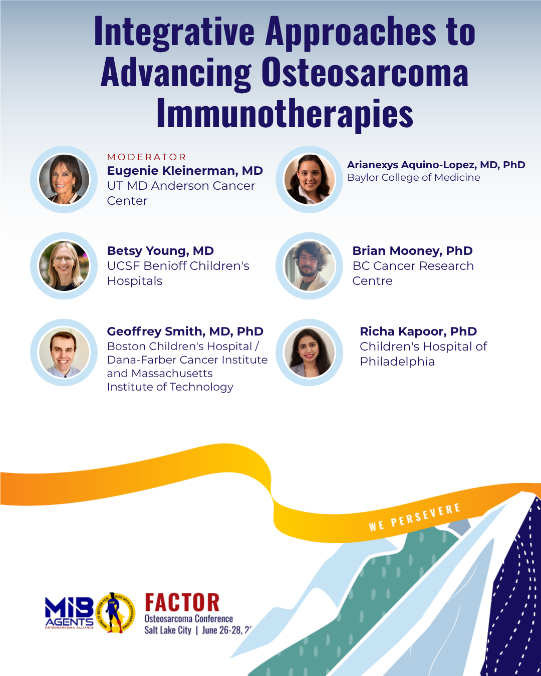
Richa Kapoor, PhD
Children's Hospital of Philadelphia
Defining the immunopeptidome of osteosarcoma nominates candidate therapeutic targets
Osteosarcoma is an aggressive childhood cancer and is often lethal despite intensive chemotherapy and radiation therapy. New cellular immunotherapies (e.g., chimer antigen receptor [CAR] T cells) have revolutionized the care of children with leukemias but as yet have not made a major impact on childhood solid cancers like osteosarcoma. One of the major barriers to progress is finding immunotherapy “targets” on the surface of cancer cells that are not present on normal cells. We developed an “immunopeptidomic” strategy to discover highly specific osteosarcoma cancer targets that are absent in normal body tissues. These are different from conventional CAR T targets (proteins on the surface of cells) as we focus on short fragments of proteins (peptides) derived from those essential for osteosarcoma and required for the cancer cell to survive. We aim to transform outcomes for children with osteosarcoma by developing novel peptide-centric CAR T cell therapies (PC-CARs) targeting these tumor-specific peptides. Our study represents a convergence of cutting-edge technologies—immunopeptidomics, and cellular therapy—to address an urgent unmet need in pediatric oncology. Our first PC-CAR targeting a PHOX2B peptide in neuroblastoma, which demonstrated remarkable pre-clinical efficacy, is set to enter clinical trials this year. For osteosarcoma, our comprehensive immunopeptidomic analysis has identified several promising peptide targets, including peptides from LRRC15, SP7 and PANX3. These targets exhibit high tumor specificity, absence in normal tissues, and are essential for cancer survival. This study is leading to the rapid development of targeted cellular therapies for osteosarcoma, laying a blueprint to follow across multiple cancer types and hence accelerating the translation of cellular therapies from bench to bedside.
Geoffrey Smith, MD, PhD
Boston Children's Hospital / Dana-Farber Cancer Institute and Massachusetts Institute of Technology
Evasion of endogenous CD8 T-cell responses in murine osteosarcoma
Forty years of progressive chemotherapy intensification in osteosarcoma (OS) has failed to improve outcomes, suggesting a new approach is needed. Immunotherapies, which target the patient’s immune system against their cancer, have revolutionized the treatment of many cancers. Unfortunately, trials of both therapies designed to boost a patient’s own immune response (termed immune checkpoint blockade) and CAR-T T cells have not yielded significant responses in OS. To understand how OS evades the immune system, we study a mouse model engineered to develop OS that has similar genetics, microscopic appearance and clinical pattern (i.e. leg tumors that spread to the lung) to human OS. Unlike models that transplant human OS into immunocompromised mice, these mice develop OS in the context of a fully functioning immune system, enabling us to study how the tumor and immune system interact. We have found that the immune system can recognize and control some OS tumors, as mice without key parts of the immune system develop OS sooner than those with intact immune systems. However, even mice with a functioning immune system eventually develop tumors. From initial studies of the immune cells found in these tumors, we propose three potential mechanisms enable immune evasion: First, the chaotic genome of OS may present so many different targets to the immune system that instead of focusing on one target, multiple small and ineffective immune responses develop. Second, the immune cells that are present in tumors may have become exhausted and no longer able to mount a response. This is a natural response to prevent autoimmunity but in this case may have been co-opted by the cancer cells. Third, the other immune cells recruited to OS may send signals to prevent an effective anti-tumor response from developing. Understanding which of these mechanisms underly immune evasion will suggest new therapies for OS and the mouse model provides a platform to rapidly test these hypotheses.
Betsy Young, MD
UCSF Benioff Children's Hospitals
cGAS-STING drives immune evasion in osteosarcoma and is therapeutically targetable via STING agonism
In cancers including osteosarcoma (OS), malfunctioning DNA replication results in a warning signal to the immune system via the cGAS-STING pathway, which we believe is modified in OS tumors to evade immune-mediated tumor killing. We have identified that half of OS patient-derived cell lines respond to cGAS-STING activation with a drug treatment, whereas the other half do not. Additionally, using RNA sequencing, we defined the specific genetic changes that occur in OS upon activation of the cGAS-STING pathway. Our studies of a STING activating drug in animal models have shown that this therapy can slow tumor growth and increase anti-tumor immunity, leading to complete regression of osteosarcoma tumors and lasting immunologic memory. Overall, we have discovered two subsets of OS with differing capacity to generate an anti-tumor immune signal and demonstrated the benefits of activating STING in patients and in preclinical models. Our proposed ongoing work will confirm the effects of STING activation, supporting the translation of this novel approach to activating immune surveillance in osteosarcoma.
Brian Mooney, PhD
BC Cancer Research Centre
Surface and global proteome profiling identifies ROR2 as an actionable immunotherapeutic target in osteosarcoma
While primary osteosarcoma is typically treated in the clinic by surgery and chemotherapy, osteosarcoma that has spread from it's primary site is a significant challenge to treat. Many adult cancers have demonstrated excellent responses to a new type of cancer treatment known as "immunotherapy", which aims to notify the patient's own immune system of the cancer and target it for destruction. Unfortunately, however, benefit to immunotherapy is somewhat lacking in pediatric solid cancers like osteosarcoma. A first step for immunotherapy is to identify a target on the surface of the cancer which can be targeted by immunotherapies. To take this first step, our team worked to generate a snapshot of the surface targets on osteosarcoma cells. One of these targets, which was very high on osteosarcoma cells but low in other cells, is known as ROR2. We went on to show that ROR2 is indeed high on osteosarcoma cells and very low in normal pediatric tissues, which is very important when designing a potential anti-cancer agent. When we selectively turned off this target in osteosarcoma cells, the cells grew slower and their ability to move was decreased. Turning this target off also stopped osteosarcoma cells from growing in the lungs of our mouse models. These trends indicate that targeting ROR2 may be beneficial to patients with osteosarcoma. We are currently working to identify and develop agents that can recognize ROR2 on the surface of osteosarcoma, and deliver anti-cancer drugs straight to the cancer. We hope that this work can help support a future clinical trial that will use immunotherapy to target ROR2 to kill osteosarcoma cells.
Arianexys Aquino-Lopez, MD, PhD
Baylor College of Medicine
Repurposing virus specific T cells as immune therapy for osteosarcoma
Osteosarcoma is a bone cancer that generally affects children and adolescents. Chemotherapy for osteosarcoma has remained the same since the 1980’s, with no improvement in survival. Furthermore, when cancer spreads to the lungs (metastatic disease), it is difficult to treat. Only between 10% to 40% of patients with lung spread survive for 5 years. Current therapies for metastatic osteosarcoma include drugs and surgery, but these often fail. To develop a better approach, I will redirect the patients’ own immune system to treat the tumor like a curable virus infection rather than a resistant cancer. When we get sick with a virus, cells from our immune system, called virus specific T cells (VSTs), eliminate unhealthy cells infected by virus. The way the immune system finds the unhealthy cells is by recognizing “viral signals” on the surface of the infected cells. I propose using a virus combination called CAdVEC to modify cancer cells and make them show these “viral signals”. This will trigger the immune cells in our body to attack and eliminate the cancer cells.
Clinical Trials
View Speakers & Abstracts.png)
Alanna Church, MD
Boston Children's / Dana-Farber Cancer Center
Focus on clinical implementation
This talk explores strategies for translating research innovations—new drugs, diagnostic tests, and lab discoveries—into clinical practice. It covers key implementation steps, regulatory pathways, and real-world integration while addressing barriers such as cost, policy constraints, stakeholder resistance, and navigating clinical systems within large healthcare organizations. Attendees will gain insights into overcoming challenges to accelerate patient access to innovations.
Brenda Weigel, MSc, MD
St. Jude Children's Research Hospital
Crucial collaborations: Key for success of pediatric oncology clinic trials
All pediatric cancers are rare diseases and require collaboration between academia and industry to successfully develop and conduct clinical trials of new therapeutic approaches. These collaborations can take many forms and be supported in many different ways. We will explore the many models for collaboration and some of the benefits challenges in developing clinical trials in these models.
Damon Reed, MD
Memorial Sloan Kettering Cancer Center
Osteosarcoma maintenance therapy with OST31-164 (OST-164-01) - Primary results
We will report the primary outcome data for the 41 patients aged 12 to 39 with osteosarcoma (bone cancer) that had recurred in the lungs who received OST31- 164 infusions every 3 weeks on AOST2121.
Kristen VanHeyst, DO
University Hospitals/Rainbow Babies and Children's Hospital
Targeting TGF-beta signaling in the tumor microenvironment as an effective therapy in osteosarcoma
Immunotherapy is an innovative field that provides novel possibilities offering hope for finding curative options for pediatric and adolescent and young adult patients. Immunotherapy has shown promise in the treatment of multiple cancers, but effective treatment responses have yet to be realized in osteosarcoma (OS). Current standard of care therapy for the treatment of OS, whether a patient has metastatic disease at the time of diagnosis or not, is the same. This standard of care has not changed in decades, and neither has the overall outcome for patients with metastatic OS or recurrent/refractory disease. The prognosis for these patients is grim. Additionally, the chemotherapeutic agents the patients receive impart caustic and sometimes devastating side effects and toxicities. For these reasons, it is imperative to advance our understanding of the OS tumor microenvironment (TME) as components of this could be targets for therapy. Transforming Growth Factor-Beta (TGF-beta) is an abundant and potent immune suppressive molecule produced by OS cells and immune cells in the TME. The presence of TGF-beta contributes to making the TME more hospitable and dampens the ability of the patient's own immune system to eradicate this tumor. TGF-beta expression is increased in the serum of patients with OS compared to healthy individuals, and high TGF-beta correlates with high grade OS and metastasis. Effective targeting of TGF-beta would therefore be a desirable therapeutic approach for treating patients. In collaboration with MedPacto, Inc, we are utilizing their first-in-class, orally available, non-toxic small molecule inhibitor, TEW-7197 (“Vactosertib”) in a Phase I/II multi-centered, multi-continental, study for pediatric, adolescent, and adult patients with Osteosarcoma. In the lab, we continue to work on ways to further exploit this potential target, combining TGF- beta inhibitor with local control.
John A. Ligon, MD
University of Florida
RNA PRIME- Harnessing tumor RNA for immunotherapy against osteosarcoma
Patients with relapsed osteosarcoma (OSA) have few effective therapeutic options. We developed a multi-lamellar RNA lipid nanoparticle (RNA-LP) incorporating autologous total tumor RNA as a novel personalized immunotherapeutic. In the first-in-human clinical trial for adults with glioblastoma, we demonstrated the capacity for this novel RNA therapeutic to generate a profound immunologic response. RNA PRIME (NCT05660408) is a “basket trial” being conducted by the Sunshine Project of the National Pediatric Cancer Foundation to treat multiple types of recurrent pediatric solid tumors and CNS tumors. The OSA arm is a first in pediatric Phase I/II trial for patients with recurrent pulmonary or unresectable OSA. The primary objective in phase I will be to demonstrate the manufacturing feasibility and safety. The primary objective in phase II will be to determine if RNA- LPs extend event-free or progression-free survival. Major eligibility criteria include age 3-39 years; evidence of recurrent OSA; able to have surgical resection or biopsy at enrolling institution (necessary for RNA isolation to generate personalized RNA- LP); and functional status at time of enrollment of >60%. Patients will enroll on one of three Arms depending on disease status at enrollment (unilateral vs. bilateral pulmonary metastatic disease vs. unresectable) and undergo surgical resection or biopsy. All arms will consist of two phases: 1) pp65 RNA-LP phase and 2) pp65/tumor mRNA RNA-LP phase, with administration spacing from an initial bi-weekly to monthly for one year. Correlative studies include peripheral immune monitoring, tumor microenvironment, and ex vivo tumor modeling of patients receiving RNA-LPs. Total accrual target is up to 18 subjects in phase I, and up to 55 subjects in phase II. Additional arms of RNA PRIME currently include an arm for patients with recurrent/progressive pediatric high grade glioma, and future arms are anticipated for additional CNS and extracranial malignancies.
CURE-OS Working Group: A Blueprint for Translating Single-Cell Discoveries into better Osteosarcoma Treatments
.png)
SARC and Other Sarcoma Mentorship and Coaching for Young Investigators in Sarcoma Research

.jpg)
Tanya Heim, MS
University of Pittsburgh and UPMC Hillman Cancer Center
Disulfiram and Enzalutamide Combination Therapy to Treat Metastatic Osteosarcoma
Using computational analyses, we previously predicted that ALDH1A1 is directly activated by androgen receptor (AR) in metastatic osteosarcoma samples. This is important because ALDH1A1 tends to be more active and abundant in osteosarcoma cells that are more aggressive, chemo-resistant, and/or in metastatic lesions. Therefore, ALDH1A1 has been a focus of our lab for quite some time and we have found that Disulfiram, an ALDH-inhibitor with low toxicity, is a promising treatment for metastatic osteosarcoma. We first aimed to validate this predicted interaction between AR and ALDH1A1 in osteosarcoma cells with varying metastatic potential by treating them with Enzalutamide, an FDA-approved AR- inhibitor, and Disulfiram separately. We initially observed that the cells with higher metastatic potential (SaOS-LM2) were more sensitive to Enzalutamide treatment. However, both cell lines displayed some sort of resistance and the effective Enzalutamide doses were much higher than what is currently deemed safe by the FDA. In this study, we tested the combination of Enzalutamide and Disulfiram treatment on osteosarcoma cells with varying metastatic potential to see if 1) the effective dose of these drugs can be reduced and 2) if the effective combination dose can prevent or reduce osteosarcoma progression and metastatic potential by specifically measuring cell proliferation (i.e. increase in the number of cells), survival (i.e. treatment resistance), and migration (i.e. metastatic potential). We found that a lower effective dose of Enzalutamide can be achieved in combination with Disulfiram to effectively reduce osteosarcoma cell proliferation and survival and that cells with higher metastatic potential are more sensitive to this combination treatment. However, we also discovered that the role of AR in metastatic osteosarcoma is not as direct and simple as once predicted and other molecules interacting with ALDH1A1 may be as important to metastatic osteosarcoma as AR.
Marika Klosowski, DVM
Colorado State University
Fibroblast Activation Protein as an Immunotherapy Biomarker and CAR T-cell Target in the Osteosarcoma Tumor Microenvironment
Osteosarcoma (OS) is the most common bone tumor affecting children and adolescent patients. While surgery and chemotherapy are effective treatments in many cases, up to 30% of OS patients sadly still die within 5 years due to spread (metastasis) of their cancer, and better treatments are urgently needed to improve the outcomes for patients with metastatic OS. OS tumors are made up of more than just cancer cells alone; they are a complex ecosystem composed of cancer cells that interacting with many other supporting non-cancerous cell types including immune cells, connective tissue fibroblasts, and blood vessel cells. This complexity makes it challenging to develop and test new treatments like immunotherapy for OS in the laboratory and this is part of the reason why effective treatments for metastatic OS are still lacking. OS is over 10 times more common in dogs than in people, and unlike laboratory mouse models, OS tumors that develop spontaneously in dogs progress very similarly to human OS and feature similar complex interactions between cancer cells and the immune system. We and others have found that a cell surface protein called fibroblast activation protein (FAP) is present on both OS cells and supporting cancer- associated fibroblasts (CAFs) in OS tumors, but not on normal body cells. Our preliminary studies also shows that OS tumors with high levels of FAP in both dogs and people have features that may make them more aggressive and resistant to treatment. We are currently testing whether a new immunotherapy regimen can be safely used in dogs and if the strategy is effective in metastatic canine OS. This approach includes genetically engineered immune cells (CAR T-cells) that use FAP as a marker to recognize and destroy cancer cells and CAFs within OS tumors, combined with two orally delivered drugs that inhibit metastasis-promoting myeloid immune cells. Together with other ongoing experiments, the information gained from examining the blood and tumor tissues of canine patients who participate in this clinical trial will also help determine whether FAP levels in OS tumors can help predict patient outcomes and responses to immunotherapy. Importantly, the drugs in this combination immunotherapy can be readily used in people, so this approach has the potential for rapid translation to human OS patient trials.
Eunice Lopez Fuentes, PhD
University of California, San Francisco
RUNX2 and FOSL1 Orchestrate Epigenetic Subtypes and Cellular Plasticity in Osteosarcoma
Our research has uncovered two subtypes of osteosarcoma, which may help doctors find better ways to treat the disease. These subtypes differ in how the cancer cells grow and respond to treatments. We found that osteosarcoma tumors can be classified into two groups, which we call EOD (Early osteoblast derived) and LOD (Late osteoblast derived). These groups are determined by how certain genes are turned on and off inside the cancer cells. EOD tumors rely on a gene called RUNX2, which is normally involved in bone development. LOD tumors, on the other hand, depend on a different gene, FOSL1, which helps cells grow and respond to stress. One important discovery is that osteosarcoma cells can switch between these two subtypes, which may explain why the cancer is so hard to treat. However, this also gives us an opportunity—if we can block the right pathways, we might be able to stop the cancer from growing. Our study also tested different treatments and found that the two subtypes respond differently to specific drugs. LOD tumors (driven by FOSL1) respond well to drugs that block a pathway called MEK, which helps cancer cells grow. EOD tumors (driven by RUNX2) are more sensitive to drugs that target a protein called AURKB, which is involved in cell division. Another key finding is that both subtypes can exist within the same tumor, making it more complex and more challenging to treat. This means that personalized treatments may be needed to target both types effectively. By identifying these two subtypes, we hope to help doctors better understand each patient’s tumor and choose treatments that are more likely to work. This research is a step toward personalized medicine for osteosarcoma, aiming to improve outcomes and give patients and families more hope for the future.
Rocio Rivera-Valentin, MD, PhD
University of Puerto Rico Comprehensive Cancer Center
Epidemiology and Molecular Determinants of Pediatric Osteosarcoma in Underserved Populations
Children with rare cancers, such as bone cancer (osteosarcoma) are often left out of scientific advancements that improve treatment for children with more common cancers (like leukemia). Because research of rare cancers is not funded as often, scientific progress is slower for these children. Indeed, while treatments for common childhood cancers have expanded to include new targeted drugs and immunotherapies, treatments for osteosarcoma have not changed in over 30 years. Osteosarcoma behaves differently in children of African or Indigenous genetic backgrounds than in children of predominantly European ancestry. Children from Black or Hispanic populations are more likely to have bone tumors that spread throughout the body, leading to poor survival outcomes, compared to other racial and ethnic groups. Until now, little is known about why these differences exist. Our group proposes to comprehensively dissect key factors that drive aggressiveness (the potential to spread) of bone tumors among Hispanic children. These factors include: (1) genetic ancestry; (2) social determinants of health (such as access to healthcare); and (3) systems inside tumors that work to shut down the immune system’s ability to fight cancer. This approach is important because it will give clinicians a first look at the big- picture elements that affect patient outcomes in these understudied populations, while simultaneously testing possible avenues for targeted therapies or immunotherapies for Hispanic children with osteosarcoma.
Busra Turgu, PhD, MSc
BC Cancer Center
Finding New Immunotherapy Targets in Osteosarcoma
Osteosarcoma (OS) is the most common bone cancer in young adults, but current treatments like chemotherapy and surgery have been ineffective, with significant side effects and a high recurrence rate. Immunotherapy, which uses the body’s own immune system to fight cancer, holds promise for long-term treatment, but it requires identifying tumor-specific targets, which remain unknown in OS. Cancer cells constantly adapt to changes in their environment, such as low oxygen levels, nutrient shortages, and oxidative stress in the tumor microenvironment. These adaptations can alter the proteins present on the cell surface, revealing new immunotherapy targets that were not discovered before. In this study, we identified proteins that are present on the surface of patient- derived osteosarcoma cells but not on normal cells. We focused on proteins that are highly specific to osteosarcoma to reduce the risk of harming healthy tissues. The goal is to use these proteins to develop CAR-T cell therapy, a personalized treatment that reprograms a patient’s immune cells to recognize and attack cancer cells more effectively. This research aims to uncover new immunotherapy targets for osteosarcoma, for more effective treatments for patients.
Yanding Zhao, PhD
Stanford University
Distinct Patterns of Chromosomal Instability Fuel Osteosarcoma Progression and Influence Patient Outcomes
Osteosarcoma (OS) is the most common primary bone malignancy in children and young adults, yet treatment has remained unchanged for four decades. With only ~175 U.S. cases annually, its rarity and extensive genomic heterogeneity have impeded biomarker discovery and targeted therapy development. Unlike many pediatric cancers, OS exhibits complex karyotypes, pervasive chromosomal instability (CIN), and diverse evolutionary trajectories. To elucidate the interplay between CIN and the tumor microenvironment in OS progression, we analyzed 91 tumor samples from 71 pediatric patients—including biopsies, resections, and metastases—using whole-genome and transcriptome sequencing. A subset was profiled with ATAC-seq, Hi-C, and IHC, with derivative cell lines available for functional studies. Over 80% of tumors harbored TP53 alterations, loss of heterozygosity, chromothripsis, kataegis, and other complex rearrangements disrupting key oncogenes and tumor suppressors. Oncogene amplification and enhancer hijacking events were validated through integrative ATAC-seq and Hi-C analysis. Replication stress at common fragile sites emerged as a key driver of CIN. Integrating rearrangement signatures with immunophenotyping, we defined four clinically distinct molecular subgroups. One subgroup exhibited homologous recombination deficiency (HRD)-associated rearrangement signatures and poor outcomes independent of immune contexture. Derivative cell lines from this group were sensitive to PARP inhibition (olaparib), revealing a potential therapeutic vulnerability. Another subgroup displayed robust immune infiltration, including myeloid-enriched populations confirmed by IHC and H&E, and showed exceptional response to chemotherapy. Together, our findings demonstrate that specific genomic rearrangement processes shape both tumor evolution and the immune landscape in OS, offering a framework for patient stratification and the development of precision therapies.
Sagar Bhattacharya, PhD | University of California, San Francisco
De Novo Design of Protease-Activated Anticancer Proteins
As the second most common cause of death in the US, cancer was expected to claim about 600,000 lives in 2022 with a mortality rate of about 1,500 a day. Despite a notable progress in treatment options including chemotherapy, radiotherapy, and immunotherapy, unfortunately, all of them suffer from side effects, which limit their efficacy as cancer therapeutics. The use of peptide drugs to treat various forms of cancer and inflammation caused by bacterial infection has recently been emerged as alternative treatment option. Despite the promises offered by bioactive peptides, very few of them could reach to the clinic in the past decade, primarily due to their off-target toxicity. Hence, design of a suitable system to deliver peptide drugs in a site-specific manner would address a major challenge in the development of anticancer and/or antimicrobial peptide drugs. Protein design from scratch has proved to be a powerful strategy to design and engineer functional proteins for a broad range of applications from catalysis to pharmaceuticals. In this work, I showed that antimicrobial peptide drugs can be caged using de novo protein design strategy and can be released by target proteases upregulated in bacterial infection for systemic delivery. I anticipate that this novel delivery system will be widely applicable to enable triggered delivery of anticancer peptides and/or small molecule drugs, beyond the antimicrobial peptides discussed here, hence expanding the repertoire of protein design to biomedical applications.
Pooja Rao, MS | Hunter College of CUNY jointly with Research Foundation of CUNY
Riluzole-Induced Radiosensitization of Osteosarcoma to Single High Dose Radiation Therapy
Osteosarcoma is an aggressive tumor of the bone mostly seen in young adults. This aggressive bone cancer is extremely resistant to traditional fractionated radiation therapy, posing substantial challenges in clinical management. Recent clinical studies indicate that single high dose radiation therapy(SDRT) as a promising alternative for treating radioresistant sarcomas. However, in osteosarcoma, delivery of very high dose of radiation to tumor site is difficult because of usually large size of primary tumors, their frequent localization in anatomically challenging regions and toxicity to the neighboring crucial tissues. Therefore, the primary challenge is to increase tumor response to SDRT to achieve safe and optimal therapeutic outcome. To address this challenge, we investigated whether combining SDRT with a drug called Riluzole could make tumors more sensitive to radiation, allowing for better treatment outcomes. We tested if Riluzole can effectively increase radiation induced DNA damage in both tumor cells and tumor blood vessels. The results from our laboratory reveal that Riluzole effectively increases sensitivity of osteosarcoma cells to radiation therapy. It increases DNA damage and apoptosis, reduces survival fraction of cells treated with SDRT, compared to SDRT alone. It also decreases the levels of vascular endothelial growth factor A (VEGFA), a key protein involved in tumor angiogenesis and radiation resistance. This novel dual targeting approach could enhance effectiveness of SDRT, while minimizing its side effects. To translate these findings into clinically relevant model, we aim to test the combination of Riluzole and SDRT on patient derived xenograft cell lines and mouse model of osteosarcoma. By demonstrating the potential of Riluzole as a radiosensitizer for osteosarcoma, the project offers a pioneering strategy to enhance the effectiveness of radiation therapy, ultimately improving survival rate in patients with this radioresistant tumor.
Marta Roman Moreno, PhD | University of California, San Francisco
Dissecting and Targeting Metastatic Drivers in Osteosarcoma
Osteosarcoma (OS) is an aggressive bone cancer that primarily affects adolescents and young adults. Although many patients are diagnosed with localized tumors based on imaging, the majority already have undetectable micrometastases at the time of diagnosis. While the standard MAP chemotherapy regimen (Methotrexate, Adriamycin, Platinum) provides long-term remission for some patients, many experience disease relapse due to the persistence of metastatic cells. This highlights the urgent need to better understand the mechanisms that enable metastatic survival and progression in order to develop more effective therapeutic strategies. To identify genes that contribute to OS metastasis, we analyzed gene expression and chromatin accessibility data from patient tumor samples and patient-derived cell lines. Using this information, we designed a focused CRISPR interference (CRISPRi) screen targeting 306 genes that were either highly expressed or exhibited open chromatin regions in metastatic samples. This screen was conducted in an orthotopic mouse model of metastatic OS, allowing us to identify genes that influence the metastatic process. Among the candidates, we identified KLF5, MEF2B, TFAP2A, and DLX6 as potential regulators of OS metastasis. We prioritized KLF5 for further study due to its potential role as a key driver of metastasis. Functional experiments using CRISPRi-mediated KLF5 knockdown demonstrated a significant reduction in primary tumor growth and improved overall survival in an OS mouse model. Additionally, RNA sequencing analysis of KLF5-knockdown cells revealed changes in gene expression and signaling pathways related to migration, further supporting its role in metastatic progression. These findings establish KLF5 as a promising therapeutic target for metastatic OS and demonstrate the utility of in vivo CRISPRi screening in identifying novel drivers of cancer progression.
Abena Peasah, MS | Stanford University with Michelle Tai, Mark Fleck, Jeehee Lee, CallanMonette, Prof. E. Alejandro Sweet-Cordero, Prof. Fan Yang
A Tissue-Engineered 3D Model for Deciphering the Role of Macrophages in Osteosarcoma Drug Responses and Biology
Osteosarcoma (OS) is an aggressive bone cancer. Despite decades of research, survival rates have not improved in nearly 40 years. One type of immune cell, the tumor-associated macrophage (TAM), is commonly found in OS tumors and has been linked to resistance to chemotherapy. This makes TAMs a promising target for new treatments. However, most studies on TAMs focus on soft tissue cancers, leaving their role in OS largely unknown. To address this gap, our research team has developed a tissue-engineered 3D OS model that mimics the bone environment. This model is built using gelatin microribbon scaffolds (Gel µRBs), which resemble the composition of bone extracellular matrix (ECM). Our team previously showed that this 3D model outperforms conventional 2D culture in mimicking OS tumor biology and drug responses in vivo. Here, we aim to leverage this 3D model to study how the interaction between TAMs and OS influences OS biology and drug response. We created the 3D co-culture model by encapsulating OS cells and macrophages together in the Gel µRBs. OS cells were labeled with luciferase to facilitate noninvasive tracking of their proliferation over time. Human monocytes were stimulated to become macrophages before being mixed into the co-culture. We then assessed the effect of 3D co-culture on OS drug responses and biology. Our results show that both OS cells and TAMs survive well in the 3D model. Interestingly, co-culture with TAMs increased OS sensitivity to the standard chemotherapy gemcitabine. At 0.01 µM , OS monoculture showed only 5% cell death, while those co-cultured with TAMs showed 9x higher death (44%). This suggests that interactions between OS cells and TAMs can change how OS tumors respond to chemotherapy. This study highlights the importance of using 3D models to better understand OS and immune cell interactions. Our findings suggest that targeting TAMs could improve chemotherapy effectiveness. Moving forward, we will expand our research to test additional drugs and analyze how TAMs influence aspects of OS biology like stemness and metastasis. This model could serve as a powerful tool for screening and discovering new treatments for osteosarcoma via targeting OS-TAM interactions.
Jeehee Lee, PhD | Stanford University
Personalized Cancer Research: Bioengineered 3D Osteosarcoma Models to Investigate Tumor Heterogeneity and Drug Responses
Bioengineered 3D Osteosarcoma models for Drug Screening and Precision Medicine Osteosarcoma is a rare and aggressive bone cancer with high genomic complexity and heterogeneity in drug responses, making treatment challenging. Traditionally, researchers have studied osteosarcoma using two-dimensional (2D) culture, which fails to mimic the complex tumor environment inside the body. To address this, this study harnessed a more advanced bioengineered 3D OS model that better mimics the extracellular matrix of OS tumors. Specifically, we seek to assess if such 3D models would better mimic drug responses in vivo than conventional 2D culture, and assess their potential for drug screening and precision medicine. Using tumor cells isolated and derived from eight osteosarcoma patients, we first created personalized 3D OS models by encapsulating them in a 3D scaffold that mimics bone matrix compositions. We then compared how the 3D OS models reacted to various drug treatments, and compared that to 2D and in vivo conditions. We found that drug response pattern of patient tumor cells grown in 3D showed a much closer match to in vivo tumor treatment outcomes, compared to 2D culture. When we screened a large panel of OS drugs across the eight patient cells, the 3D models displayed a wider range of drug responses than 2D, revealing important differences in drug effectiveness across patients. Additionally, tumors in the 3D model produced more extracellular matrix (ECM), an important aspect of tumor niche that can influence drug response. These findings highlight the potential of 3D bioengineered OS models as a powerful tool to study the differences in OS tumor across patients, and screen drugs that could better predict how patients might respond to potential treatments. By improving drug testing accuracy with reduced time and cost, such 3D bioengineered models could pave the way for more personalized and effective treatments for OS patients in the future.
Lu Xie, MD | Peking University People's Hospital
Chemotherapy-Induced Thrombocytopenia in Patients With Primary Bone Sarcomas: Current Status and Approach to Management: A LargeScale Real-World Analysis
Background: Multi-agent chemotherapy is an integral part and first-line systemic treatment for primary bone sarcomas, but it is limited by myelosuppression and thrombocytopenia, resulting in chemotherapy delays or dose reduction. The study aimed to record the changes in platelet counts and investigate the demographics and influencing factors of platelet counts after chemotherapy, identifying the clinical risk factors for thrombocytopenia. Methods: This was a single-center, retrospective analysis conducted at Peking University People’s Hospital, China. A total of 1,000 chemotherapy courses in 462 patients aged 2–67 were reviewed. Platelet counts ˂20 x109/L or 50 x109/L were considered as clinically notable study events. Results: Following first- or second-line chemotherapies, we explored the dynamic curve of platelet changes. A total of 65 (14%) and 191 (41%) out of 462 patients had platelet counts ˂20 x109/L and 50 x109/L, respectively. The number of previous lines of treatment (P =0.0161) and chemotherapy regimens (P = 0.0000) were the most significant risk factors of thrombocytopenia. PA/A (cisplatin and adriamycin or single adriamycin) and IE/IFO ifosfamide and etoposide or single ifosfamide) were the chemotherapy regimens that had the most significant impact on platelet levels. Spider plots of platelet counts showed that the IE/IFO regimen resulted in earlier platelet nadirs. The duration of platelet-elevating drugs (avatrombopag, herombopag, recombinant human TPO and interleukin 11) also affected platelet counts after chemotherapy. A longer duration of medication was associated with better platelet level, which could avoid thrombocytopenia (≥5 days versus <5 days, P = 0.0000). Conclusion: Thrombocytopenia is a frequent adverse event occurring during chemotherapy in patients with primary bone sarcomas. Patients with more lines of previous chemotherapy and those who experienced IE/IFO or PA/A chemotherapy regimens may require closer follow-up with early initiation of platelet-boosting therapy, which may help to prevent platelet reduction from affecting the efficacy of chemotherapy.
Lu Xie, MD | Peking University People's Hospital
B7-H3 as a Prognostic Biomarker in Osteosarcoma: Insights into Clinical Outcomes
B7-H3, an immune checkpoint molecule, plays a critical role in regulating tumor immune evasion and progression. Increasing evidence suggests that soluble B7-H3 (sB7-H3) levels are associated with unfavorable outcomes in patients with malignant tumors. In this study, we quantified sB7-H3 expression detecting its changes before and after chemotherapy in 100 newly diagnosed osteosarcoma (OS) patients using ELISA and analyzed its correlation with clinical characteristics. We found that the change rate of sB7-H3 was significantly higher in patients with disease progression and low necrosis rates compared to those without progression and with high necrosis rates (P < 0.05). Furthermore, we also evaluated the diagnostic utility of sB7-H3 in predicting progression-free survival (PFS) in OS patients and found that sB7-H3 change rate exceeding 11.5% was associated with poor prognosis. These findings indicate that serum sB7-H3 serves as a valuable biomarker in OS, and high levels of sB7-H3 change rates are associated with poor clinical outcomes and disease progression.